Venous-sinus-techniques: Atlanto-occipital release
venous-sinus-techniques: Atlanto-occipital release:
|
Contraindications
|
|
Fractured dens of axis |
|
danger of intracranial bleeding (haemorrhage) |
|
Fracture in base of skull |
|
Effect
|
|
Release of the atlanto-occipital joint |
|
Release in the area of jugular foramen |
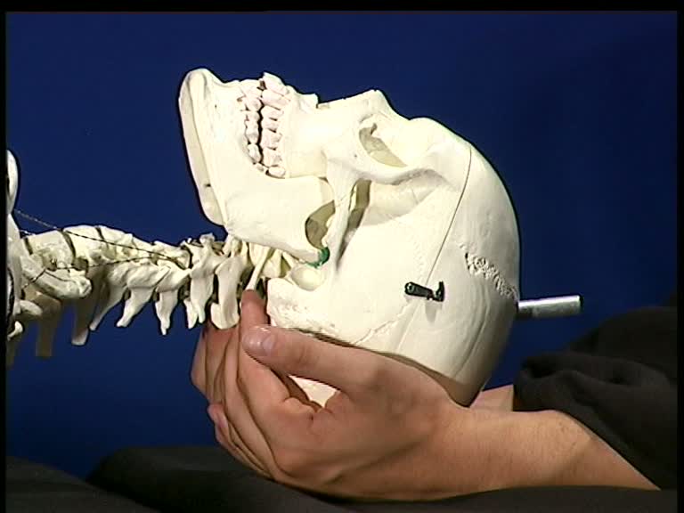
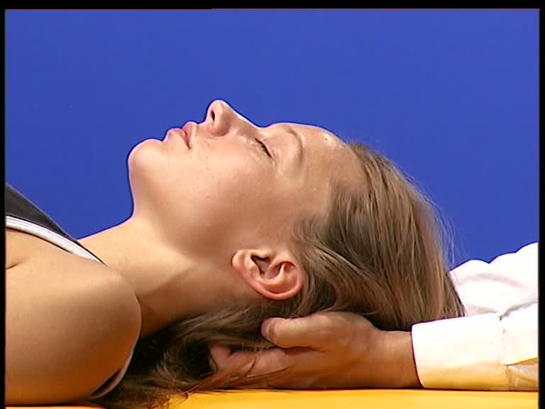
 -Both hands lie under the occipital bone
-The occipital squama lies in the palms of the hands.
-The fingers are placed in right angles, so that they’re pointing strictly anterior.
-The finger tips lie directly underneath the lower occipital edge (rim).
-Through the weight of the head, the neck muscles relax increasingly using the fingers as a lever. No additional pressure is given with the fingers.
- If the fingers start slanting due to the relaxation of the neck muscles, they are repeatedly placed at right angles.
 - With increased relaxation of the neck muscles, one can start feeling the bony arch of the atlas
- Towards the end of the treatment, the head does not lie in the palms of the hands anymore, but is only supported by the fingers on the atlas.
- After the relaxation of the neck muscles the occipital condyles are released from the atlas. The middle fingers fixate the arch of the atlas while the ring fingers and little fingers gently pull the occiput cranial.
- After that, one does a transverse decompression of the occipital condyles. For that, the fingers are placed in a 45° angle toward the foramen magnum. For an intraosseal decompression, the therapist brings the elbows closer together, causing the fingers at the occipital condyles to move apart.


